Knee Muscle Anatomy Mri - Mri anatomy of knee Dr. Muhammad Bin Zulfiqar / When interpreting the proton density images it.. Knee muscles need to have both good strength and flexibility. Tibial tuberosity with distal patella tendon insertion. An mri of the knee of a healthy subject was performed in the 3 planes of space (coronal, axial, sagittal) commonly used in osteoarticular imaging, with two weightings most commonly used to explore the musculoskeletal pathology of the knee: Magnetic resonance imaging (mri) interpretation of the knee is often a daunting challenge to the student or physician in training. This mri knee cross sectional anatomy tool is absolutely free to use.
The muscles of the knee include the quadriceps, hamstrings, and the muscles of the calf. Knee muscle anatomy axial mri : This mri knee cross sectional anatomy tool is absolutely free to use. Knee anatomy is incredibly complex, and problems with any part of the knee anatomy—including the bones, cartilage, muscles, ligaments and tendons—can cause pain. 4, infrapatellar fat pad of hoffa.
Example of MRI slice mid-thigh with the knee extensors and ... from www.researchgate.net Prescribe sagittal plane off axial images with line parallel to bony glenoid. These motions of the knee allow the body to perform such important movements as walking, running, kicking, and jumping. Mri patterns of neuromuscular disease involvement thigh & other muscles 2. The muscles of the knee include the quadriceps, hamstrings, and the muscles of the calf. Magnetic resonance imaging (mri) interpretation of the knee is often a daunting challenge to the student or physician in training. This mri knee cross sectional anatomy tool is absolutely free to use. Knee mri (an approach) dr dai roberts and dr joachim feger et al. Magnetic resonance imaging is particularly well suited for the medical evaluation of the musculoskeletal (msk) system including the knee, shoulder, ankle, wrist and elbow.
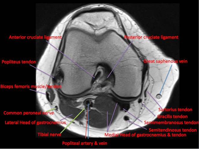
The knee joint is a synovial joint which connects the femur thigh bone the longest bone in the body to the tibia shin bone.
Atlas of knee mri anatomy. An mri of the knee of a healthy subject was performed in the 3 planes of space (coronal, axial, sagittal) commonly used in osteoarticular imaging, with two weightings most commonly used to explore the musculoskeletal pathology of the knee: Song, uc san francisco msiv gillian lieberman md. Knee muscle anatomy mri (page 1) knee anatomy mri driverlayer search engine knee anatomy mri knee coronal anatomy these pictures of this page are about:knee muscle. Both the pronounced accuracy of the mri and the high prevalence of knee disorders, makes the knee mri the most frequently ordered imaging procedure of the musculoskeletal system. The muscles that affect the knee's movement run along the thigh and calf. Louis, usa and the rijnland hospital in leiderdorp, the netherlands. Magnetic resonance imaging is particularly well suited for the medical evaluation of the musculoskeletal (msk) system including the knee, shoulder, ankle, wrist and elbow. Knee mri (an approach) dr dai roberts and dr joachim feger et al. These muscles work in groups to flex, extend and stabilize the knee joint. Can also generate proton density images. There is a flat area of tendon originating from the knee. Use the checklist to quiz yourself.
An mri of the knee of a healthy subject was performed in the 3 planes of space (coronal, axial, sagittal) commonly used in osteoarticular imaging, with two weightings most commonly used to explore the musculoskeletal pathology of the knee: These muscles work in groups to flex extend and stabilize the knee joint. From superficial to deep includes the pes anserinus tendons, semimembranosus tendon, tibial collateral ligament, meniscofemoral and meniscotibial ligaments, and the medial meniscus. These motions of the knee allow the body to perform such important movements as walking, running, kicking, and jumping. Coronal anatomy of the knee.
Knee Muscle Anatomy Mri : Atlas Of Knee Mri Anatomy W ... from epos.myesr.org Radiology department of the washington university school of medicine, st. The muscles that affect the knee's movement run along the thigh and calf. In this presentation mri anatomy biceps femoris muscle. Use the checklist to quiz yourself. 12 photos of the knee muscle anatomy mri. Injuries such as anterior cruciate ligament, meniscus and rotator cuff tears are all easily diagnosed when there is a firm understanding and knowledge of human anatomy. Radiology imaging medical imaging subscapularis muscle shoulder anatomy bicep tendonitis mri brain shoulder rehab rotator cuff tear anatomy this. Knee muscle anatomy axial mri :
Knee anatomy is incredibly complex, and problems with any part of the knee anatomy—including the bones, cartilage, muscles, ligaments and tendons—can cause pain.
These motions of the knee allow the body to perform such important movements as walking, running, kicking, and jumping. Knee muscles need to have both good strength and flexibility. Radiology imaging medical imaging subscapularis muscle shoulder anatomy bicep tendonitis mri brain shoulder rehab rotator cuff tear anatomy this. This mri knee cross sectional anatomy tool is absolutely free to use. Knee mri (an approach) dr dai roberts and dr joachim feger et al. The images may also help physicians to distinguish normal, healthy tissues from dead tissues(2). Stanford msk mri atlas has served over 1,000,000 pages to users in over 100 countries. T2w axial fat sat 1. Atlas of knee mri anatomy. Naturally, in order to assess pathologic knee imaging, it is necessary to know the appearance of a normal knee mri. Use the checklist to quiz yourself. This mri knee sagittal cross sectional anatomy tool is absolutely free to use. Knee muscles need to have both good strength and flexibility.
From superficial to deep includes the pes anserinus tendons, semimembranosus tendon, tibial collateral ligament, meniscofemoral and meniscotibial ligaments, and the medial meniscus. An mri of the knee of a healthy subject was performed in the 3 planes of space (coronal, axial, sagittal) commonly used in osteoarticular imaging, with two weightings most commonly used to explore the musculoskeletal pathology of the knee: Tibial tuberosity with distal patella tendon insertion. Involved early gray = muscle: 12 photos of the knee muscle anatomy mri.
View Image from www.nrronline.org Find out about how the different muscles of the knee work and how they get injured. Coronal anatomy of the knee. Song, uc san francisco msiv gillian lieberman md. Use the checklist to quiz yourself. These muscles work in groups to flex, extend and stabilize the knee joint. Naturally, in order to assess pathologic knee imaging, it is necessary to know the appearance of a normal knee mri. Learn about mri anatomy with free interactive flashcards. Can also generate proton density images.
From superficial to deep includes the pes anserinus tendons, semimembranosus tendon, tibial collateral ligament, meniscofemoral and meniscotibial ligaments, and the medial meniscus.
Doctors may recommend a knee mri if a patient experiences the following(3): Find out how the different structures fit together in our knee diagram the knee joint is the largest and one of the most complex joints in the human body. Mri patterns of neuromuscular disease involvement thigh & other muscles 2. Radiology imaging medical imaging subscapularis muscle shoulder anatomy bicep tendonitis mri brain shoulder rehab rotator cuff tear anatomy this. Coronal anatomy of the knee. Learn about mri anatomy with free interactive flashcards. This article is based on a presentation given by david rubin and adapted for the radiology assistant by robin smithuis. In one investigation, depicted only on the proton density weighted images. Abnormal anatomy with normal signal, i.e. Medical images from an mri allow medical professionals to distinguish body tissues, including the meniscus (shock absorbers in the knee), cartilage, tendons, and ligaments. Serves as a paid consultant to or is an employee of conformis inc.; Knee mri (an approach) dr dai roberts and dr joachim feger et al. 4, infrapatellar fat pad of hoffa.